

X-Rays: What Should You Know?
An X-ray is a fast, painless imaging test that takes pictures of bones, joints, and soft tissues using safe, controlled amounts of radiation. At Jangra Diagnostics Rohtak, our digital X-ray suite delivers instant high-contrast imaging under senior radiologist supervision.
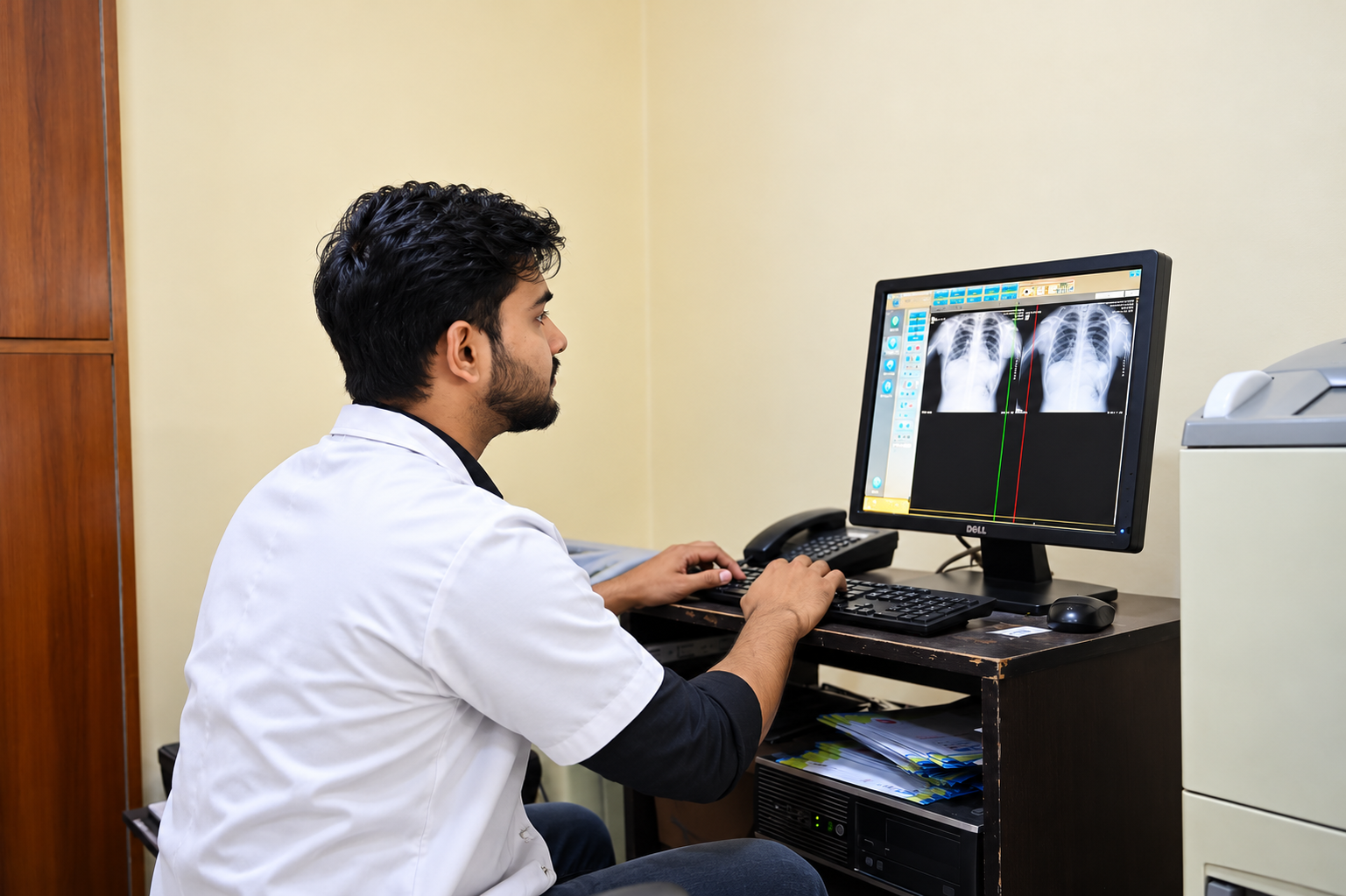
Clear Bone and Soft Tissue Imaging for Accurate Diagnostics
X-rays use safe, controlled amounts of radiation to create sharp diagnostic pictures of bones and soft tissues. These images help healthcare providers diagnose fractures, joint dislocations, chest infections, and internal structural problems, guiding prompt and effective clinical treatments.
Common Uses: Why Does a Doctor Order an X-Ray?
Fast, accurate detection of fractures, hairline cracks, dislocations, and bone trauma after injury.
Identifying underlying joint effusion, arthritis, degenerative changes, or soft-tissue swelling.
Locating radio-opaque foreign bodies lodged in soft tissues, respiratory passages, or the digestive tract.
Evaluating spinal alignment (scoliosis, lordosis), joint spaces, bone density loss, and congenital deformities.
Pre-surgical planning, verifying orthopedic implant alignment, and tracking bone healing post-casting or surgery.
Chest X-rays for pneumonia, tuberculosis, pulmonary infections, cardiomegaly, and routine medical fitness.
What are the Types of X-Ray Studies?
Several types of X-rays capture images of different anatomical areas:
Bone & Skeletal X-Rays
High-contrast digital radiographs of extremities, skull, pelvis, and joints (knee, hip, shoulder, ankle, wrist) to detect fractures, bone infections, or arthritis.
Chest X-Ray (PA & AP Views)
Standard non-invasive imaging evaluating lung fields, airways, heart silhouette, mediastinum, and chest wall.
Abdominal & KUB X-Rays
Visualizing bowel gas patterns, intestinal obstructions, fluid levels, and radio-opaque kidney or urinary bladder calculi.
Spine Investigations
Cervical, thoracic, and lumbosacral spine series (AP, Lateral, Flexion/Extension) assessing disc spaces, spondylosis, and vertebral alignment.
Digital Dental X-Rays (RVG)
High-resolution intraoral radiographs providing detailed views of tooth roots, bone levels, and hidden interproximal decay.
Special Contrast Studies & HSG
Specialized investigations including Hysterosalpingography (HSG for uterine and fallopian tube patency) and GI contrast examinations.
What is an X-Ray with Contrast Material?
Some special X-rays utilize a contrast material (also called contrast agent or dye) to highlight specific organs, blood vessels, or hollow tracts with greater clarity. Depending on the test, contrast dye may be administered:
Taken by mouth as a liquid drink (e.g., Barium swallow / meal)
Injected through an IV line into a vein (e.g., IVP renal studies)
Administered via enema for lower gastrointestinal imaging
How Do I Prepare for an X-Ray?
Radiation from an X-ray can harm your developing fetus. If you are pregnant or think you might be pregnant, you must inform your doctor and our radiographer before getting an X-ray. Alternative non-radiation imaging (such as ultrasound) will be recommended whenever possible.
What to Expect After Your X-Ray & Safety Risks
After an X-ray with contrast dye: Drink plenty of water throughout the day to flush the contrast material naturally from your body. A small number of patients may experience mild side effects such as nausea, temporary stomach cramps, mild diarrhea, or headaches.
Allergic Reactions (Rare): Rarely, an allergic reaction to contrast dye can occur. Individuals with pre-existing allergies or asthma have a slightly higher risk. Call your provider right away if you develop hives, itching, or breathing difficulty.
Radiation Risk Overview: Although X-rays use radiation, modern digital systems utilize minimal radiation doses tailored specifically to the body part (ALARA - As Low As Reasonably Achievable). There is a very low risk of overexposure, and digital X-rays are universally regarded as safe and highly effective for patients of all ages.
Book a Digital X-Ray
Walk-in X-rays and specialized investigations available Monday to Sunday with minimal wait times.