

Ultrasound (Sonography): What Should You Know?
Ultrasound uses high-frequency sound waves to create real-time pictures of internal organs, soft tissues, and blood vessels with zero radiation. Conducted and interpreted by Dr. Krishan Jangra (MBBS, DMRD, DNB) at Jangra Diagnostics, Rohtak.
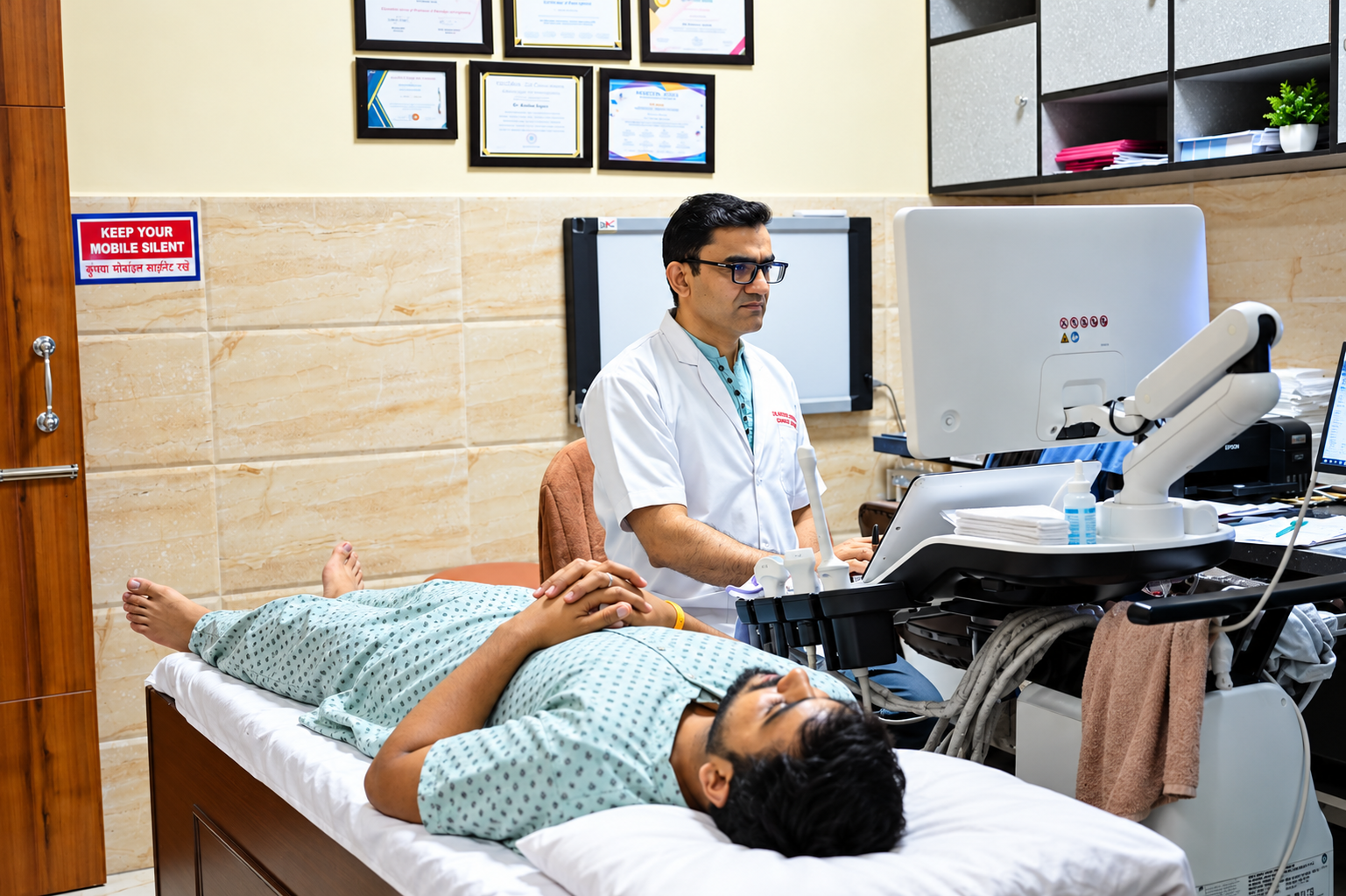
Clear Visualisation of Soft Tissues Without Radiation
Ultrasound (also known as sonography or ultrasonography) enables healthcare providers to examine internal organs, muscles, tendons, and blood vessels in real time without making any cuts or incisions. Because it uses sound waves rather than X-rays, it carries no radiation risk and is completely safe during pregnancy.
What are the Different Kinds of Ultrasounds?
Ultrasound imaging generally falls into three clinical categories:
Diagnostic Ultrasound
Used to view internal organs and soft tissues to investigate unexplained pain, palpated lumps or masses, or abnormal laboratory findings.
- Whole abdomen and pelvic organs
- Kidney, ureter and urinary bladder (KUB)
- Gallbladder, liver, spleen and pancreas
- Thyroid, neck, scrotum, and breast sonomammography
Pregnancy Ultrasound
Safe, non-invasive imaging used to monitor every milestone of maternal-fetal development throughout gestation.
- Confirming viable pregnancy & gestational age
- Evaluating fetal heartbeat and real-time movement
- Monitoring amniotic fluid levels & placental location
- Targeted screening for congenital conditions
Ultrasound Guidance for Procedures
Real-time acoustic visualization allowing clinicians to guide needles and instruments with pinpoint clinical accuracy.
- Fine Needle Aspiration Cytology (FNAC) & biopsy
- Drainage of cysts, abscesses, or fluid collections
- Organ sampling (liver, kidney, prostate)
- Embryo transfer (IVF), nerve blocks & IUD verification
When to Get a Pregnancy Ultrasound Done
While your obstetrician will customize your scan schedule based on your individual health, the following four scans form the standard clinical framework during pregnancy:
Early Pregnancy Viability Scan
Confirms viability, establishes accurate gestational age, determines the number of fetuses (single vs. multiple), and checks implantation site.
NT/NB Scan & First Trimester Screening
Measures the nuchal translucency (fluid space at the back of the fetus's neck) and checks the presence or absence of the nasal bone. Crucial for early detection of Down syndrome and other congenital heart, abdominal, and skeletal conditions. Frequently paired with the Double Marker blood test.
Level-II Scan (Targeted Anomaly Scan)
Thorough anatomical survey to evaluate the structural integrity of the fetal brain, spine, heart, kidneys, limbs, facial features, and placenta.
Color Doppler & Fetal Growth Scan
Assesses fetal umbilical blood flow, placental perfusion, and intracranial/cardiac hemodynamics, alongside fetal weight and amniotic fluid volume.
Difference Between 2D, 3D, and 4D Ultrasound
Produces flat, outline-based anatomical slices. Essential for medical diagnostics and biometry measurements of organs and fetus.
Combines multiple sound wave angles to show realistic surface depth, allowing visualization of facial features, hands, and feet.
Shows 3D images rendered in streaming real-time video, capturing fetal yawning, kicking, or subtle facial gestures.
What Conditions Can Be Detected by Ultrasound?
Ultrasound assists clinicians in identifying a wide variety of pathological, structural, and physiological conditions:
Everything You Need to Know Before Your Scan
Q:How do I prepare for an ultrasound?
Preparation depends on the specific exam. Many ultrasounds require no preparation at all. For abdominal ultrasounds, you may need to fast (refrain from food or drink except water) for 4–6 hours before your scan. For pelvic, early pregnancy, or KUB ultrasounds, you will be asked to drink 3–4 glasses of water 1 hour prior to ensure a comfortably full bladder for clear acoustic transmission.
Q:Is an ultrasound painful?
External ultrasounds (performed over the skin with warm conductive gel) are completely painless. You will feel slight pressure from the transducer, but you won't feel the sound waves. Having a full bladder may cause mild pressure. Internal ultrasounds (such as transvaginal scans for early pregnancy or gynecology) may cause slight temporary discomfort, but they do not cause sharp pain.
Q:Are ultrasounds safe? Does it use radiation?
Yes. Ultrasound imaging is completely safe and uses non-ionizing high-frequency sound waves—it involves zero radiation. It has no harmful side effects on mothers or developing fetuses. All ultrasounds at Jangra Diagnostics are conducted with medical-grade equipment under ALARA principles.
Q:What is the difference between 2D, 3D, and 4D ultrasound?
Traditional 2D ultrasound produces flat, cross-sectional grayscale images that radiologists use for clinical measurements. 3D ultrasound combines multiple slices into a realistic volumetric surface rendering (showing facial contours, fingers, and toes). 4D ultrasound is a 3D scan in live motion. In line with FDA guidance, 3D/4D scans should always be conducted for medical indications under radiologist supervision rather than for non-medical entertainment.
Q:Who performs an ultrasound at Jangra Diagnostics?
Ultrasounds require advanced clinical training to interpret correctly. At Jangra Diagnostics, all scans are personally performed and interpreted by qualified Consultant Radiologists (Dr. Krishan Jangra - MBBS, DMRD, DNB, Ex-Senior Resident PGIMS Rohtak), ensuring strict adherence to diagnostic accuracy and ethical standards.
Book Your Ultrasound Scan
Prior appointment ensures zero waiting time. Urgent & walk-in diagnostic cases are also accommodated promptly.